Portland, Maine-based medical physicist. Research Instructor of Radiology at Stony Brook Medicine interested in contrast-enhanced breast imaging, diagnostic imaging, art, design and beautiful ideas.
2️⃣0️⃣1️⃣2️⃣🏃🏻♂️2️⃣0️⃣2️⃣1️⃣
In 2012, I trained for my first marathon while studying for Part 1 of the American Board of Radiology Medical Physicist certifying exam. Last weekend, in recognition and celebration of successfully completing the Part 3 Oral exam in May (and because I like numerical anagrams), I ran the same marathon as a Board-certified Medical Physicist. Proud and privileged to push my mind, body and spirit to the limits. (at Maine Marathon) https://www.instagram.com/p/CU0DZe9L5PlW_gh-l7j2-juVcm2uz_wLJwxNvQ0/?utm_medium=tumblr
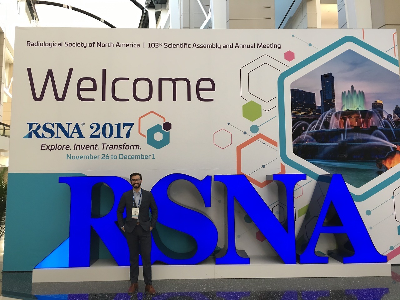
I had a great time presenting my abstracts on minimal compression and angular range in digital breast tomosynthesis at this year’s RSNA. Thanks for all of the feedback and for a great conference!
Dual-energy contrast-enhanced imaging is being investigated as a tool to identify and localize angiogenesis in the breast, a possible indicator of malignant tumors. This imaging technique requires that x-ray images are acquired at energies above the k-shell binding energy of an appropriate radiocontrast agent. Iodinated contrast agents are commonly used for vascular imaging, and require x-ray energies greater than 33 keV. Conventional direct conversion amorphous selenium (a-Se) flat panel imagers for digital mammography show suboptimal absorption efficiencies at these higher energies.
Methods
We use spatial-frequency domain image quality metrics to evaluate the performance of a prototype direct conversion flat panel imager with a thicker a-Se layer, specifically fabricated for dual-energy contrast-enhanced breast imaging. Imaging performance was evaluated in a prototype digital breast tomosynthesis (DBT) system. The spatial resolution, noise characteristics, detective quantum efficiency and temporal performance of the detector were evaluated for dual-energy imaging for both conventional full-field digital mammography (FFDM) and DBT.
Results
The zero-frequency detective quantum efficiency of the prototype detector is improved by approximately 20% over the conventional detector for higher-energy beams required for imaging with iodinated contrast agents. The effect of oblique entry of x-rays on spatial resolution does increase with increasing photoconductor thickness, specifically for the most oblique views of a DBT scan. Degradation of spatial resolution due to focal spot motion was also observed. Temporal performance was found to be comparable to conventional mammographic detectors.
Conclusions
Increasing the a-Se thickness in direct conversion flat panel imagers results in better performance for dual-energy contrast-enhanced breast imaging. The reduction in spatial resolution due to oblique entry of x-rays is appreciable in the most extreme clinically relevant cases, but may not profoundly affect reconstructed images due to the algorithms and filters employed. Degradation to projection domain spatial resolution is thus outweighed by the improvement in detective quantum efficiency for high-energy x-rays.